- Parents Home
- Para Padres
- A to Z Dictionary
- Allergy Center
- Asthma
- Cancer
- Diabetes
- Diseases & Conditions
- Doctors & Hospitals
- Emotions & Behavior
- First Aid & Safety
- Flu (Influenza)
- Food Allergies
- General Health
- Growth & Development
- Heart Health & Conditions
- Homework Help Center
- Infections
- Newborn Care
- Nutrition & Fitness
- Play & Learn
- Pregnancy Center
- Preventing Premature Birth
- Q&A
- School & Family Life
- Sports Medicine
- Teens Home
- Para Adolescentes
- Asthma
- Be Your Best Self
- Body & Skin Care
- Cancer
- Diabetes
- Diseases & Conditions
- Drugs & Alcohol
- Flu (Influenza)
- Homework Help
- Infections
- Managing Your Weight
- Medical Care 101
- Mental Health
- Nutrition & Fitness
- Q&A
- Safety & First Aid
- School, Jobs, & Friends
- Sexual Health
- Sports Medicine
- Stress & Coping
X-Ray Exam: Chest
What's an X-Ray?
An X-ray is a safe and painless test that uses a small amount of radiation to make an image of bones, organs, and other parts of the body.
The X-ray image is black and white. Dense body parts, such as bones, block the passage of the X-ray beam through the body. These look white on the X-ray image. Softer body tissues, such as the skin and muscles, allow the X-ray beams to pass through them. They look darker on the image.
X-rays are commonly done in doctors’ offices, radiology departments, imaging centers, and dentists’ offices.
What's a Chest X-Ray?
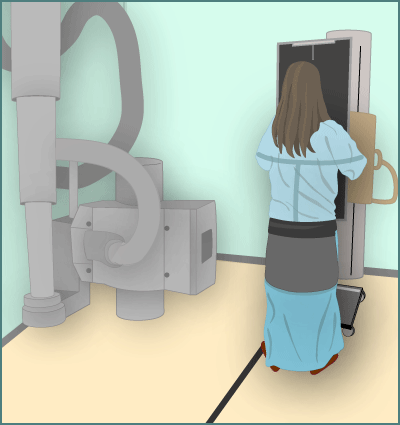
In a chest X-ray, an X-ray machine sends a beam of radiation through the chest, and an image is recorded on special film or a computer. This image includes organs and structures such as the heart, lungs, large blood vessels, diaphragm, part of the airway, the upper spine, ribs, collarbone, and breastbone.
Usually, the X-ray technician will take pictures of the chest:
- from the back of the chest (if the child is old enough to stand up for the X-ray)
- from the side
For younger children, the technician will take pictures from the front of the chest and the side. Sometimes, other picture views also are taken.
Chest X-rays can be done while a child is standing, sitting, or lying down. They should stay still for 2–3 seconds while each X-ray is taken so the images are clear. If an image is blurred, the X-ray technician might take another one.

Why Are Chest X-Rays Done?
A chest X-ray can help doctors find the cause of a cough, shortness of breath, or chest pain. It can detect signs of pneumonia, a collapsed lung, heart problems (such as an enlarged heart), and broken ribs or lung damage after an injury.
Chest X-rays can show a swallowed foreign object (such as a coin). They can also help confirm that medical tubes are in the right locations in organs such as the stomach.
What if I Have Questions?
If you have questions about the chest X-ray or what the results mean, talk to your doctor.