Corporate Circle Members
40 Exchange Place, Suite 1902, New York, NY 10005
Phone
917.746.8300
Members Only
877.662.7627
© 2020 NAPNAP, Inc. All rights Reserved
A computed tomography scan (CT scan), also called computed axial tomography scan (CAT scan), is a type of imaging test. It uses computers and a rotating X-ray machine to take cross-sectional pictures of the body. CT scans give doctors more detailed images than X-rays can provide. Unlike X-rays, they can show organs, soft tissues, and blood vessels in addition to bones.
CT scans are painless. A CT scan involves more exposure to radiation than a regular X-ray does, but the risk is small.
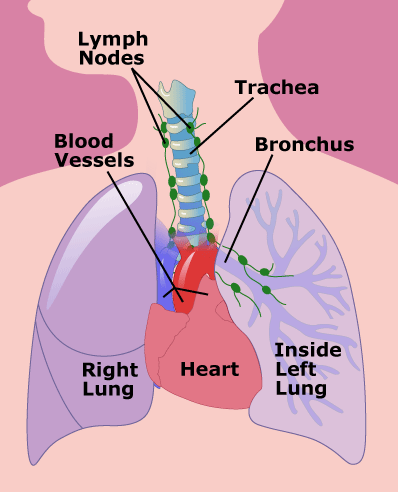
A chest CT scan uses a special X-ray machine to take pictures of a patient's lungs, heart, blood vessels, airway passages, ribs, and lymph nodes.
A person getting a CT scan lies on a table. A pillow and straps hold them in place to prevent movement that would result in a blurry image. The donut-shaped machine circles the body, taking pictures to provide cross-sections of the chest from various angles. These pictures are sent to a computer that records the images. It also can put them together to form 3D images.
A chest CT scan can find signs of inflammation, infection, injury or disease of the lungs, breathing passages (bronchi), heart, major blood vessels, , and esophagus.
If you have questions about the chest CT scan or what the test results mean, talk to your doctor.


© 1995- The Nemours Foundation. KidsHealth® is a registered trademark of The Nemours Foundation. All rights reserved.
Images sourced by The Nemours Foundation and Getty Images.
New UsersCreate a User AccountBecome a Member |
Forgot PasswordForgot your password? Click here |
