Your Heart & Circulatory System
The Heart
What Is the Heart and What Does It Do?
The heart is an organ that’s a pump, usually beating about 60 to 100 times per minute. With each heartbeat, the heart sends blood throughout your body, carrying oxygen to every cell. After delivering the oxygen, the blood returns to the heart. The heart then sends the blood to the lungs to pick up more oxygen. This cycle repeats over and over again.
What Are the Parts of the Heart?
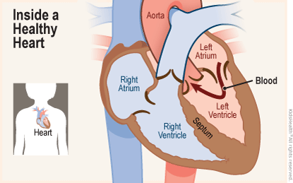
Heart Chambers
The heart has four chambers — two on the top and two on the bottom:
- The two bottom chambers are the right ventricle and the left ventricle. These pump blood out of the heart. A wall called the interventricular septum is between the two ventricles.
- The two top chambers are the right atrium and the left atrium. They receive the blood entering the heart. A wall called the interatrial septum is between the atria.
Heart Valves
Parts of the heart called valves keep blood moving through the heart in the right direction. The atria are separated from the ventricles by the atrioventricular valves:
- The tricuspid valve separates the right atrium from the right ventricle.
- The mitral valve separates the left atrium from the left ventricle.
Two valves also separate the ventricles from the large blood vessels that carry blood leaving the heart:
- The pulmonic valve is between the right ventricle and the pulmonary artery, which carries blood to the lungs.
- The aortic valve is between the left ventricle and the aorta, which carries blood to the body.
How Does the Heart Beat?
The heart gets messages from the body that tell it when to pump more or less blood depending on a person's needs. For example, when you're sleeping, it pumps just enough to provide for the lower amounts of oxygen your body needs when resting. But when you're exercising, the heart pumps faster so that your muscles get more oxygen and can work harder.
The way the heart beats is controlled by a system of electrical signals in the heart.
Sinus and Atrioventricular Nodes
The sinus node, or sinoatrial node, is a small area of tissue in the wall of the right atrium. It sends out an electrical signal to start the contracting (pumping) of the heart muscle. This node is called the pacemaker of the heart because it sets the rate of the heartbeat and causes the rest of the heart to contract in its rhythm.
These electrical impulses make the atria contract first. Then the impulses travel down to the atrioventricular node, or AV node, which acts as a kind of relay station. From here, the electrical signal travels through the right and left ventricles, making them contract.
Systole and Diastole
One complete heartbeat is made up of two phases:
Systole
The first phase is called systole (pronounced: SISS-tuh-lee). This is when the ventricles contract and pump blood into the aorta and pulmonary artery. During systole, the atrioventricular valves close, creating the first sound — the lub — of a heartbeat. When the atrioventricular valves close, this keeps the blood from going back up into the atria.
During this time, the aortic and pulmonary valves are open to let blood into the aorta and pulmonary artery. When the ventricles finish contracting, the aortic and pulmonary valves close to prevent blood from flowing back into the ventricles. These valves closing is what creates the second sound — the dub — of a heartbeat.
Diastole
The second phase is called diastole (pronounced: die-AS-tuh-lee). This is when the atrioventricular valves open and the ventricles relax. This allows the ventricles to fill with blood from the atria and get ready for the next heartbeat.
The Circulatory System
What Is the Circulatory System and What Does It Do?
The circulatory system is made up of your heart and three main kinds of blood vessels: arteries, veins, and capillaries. The system moves blood in these blood vessels throughout the body.
The circulatory system carries oxygen, nutrients, and hormones to cells, and removes waste products, like carbon dioxide. These roadways travel in one direction only, to keep things going where they should. Arteries carry blood away from the heart, and veins carry blood back to the heart. Capillaries are tiny blood vessels that connect very small arteries to very small veins.
What Are the Parts of the Circulatory System?
Two pathways come from the heart: pulmonary circulation and systemic circulation.
Pulmonary Circulation
The pulmonary circulation is a short loop from the heart to the lungs and back again.
The pulmonary artery is a big artery that comes from the heart. It splits into two main branches and brings blood from the heart to the lungs. At the lungs, the blood picks up oxygen and drops off carbon dioxide. The blood then returns to the heart through the pulmonary veins.
Systemic Circulation
The systemic circulation carries blood from the heart to all the other parts of the body and back again.
Blood that returns to the heart after pulmonary circulation has picked up lots of oxygen from the lungs. So it can now go out to the body. The aorta is the largest artery, and it leaves the heart carrying the oxygen-rich blood. Branches from the aorta send blood to the muscles of the heart itself, as well as all other parts of the body. Like a tree, the branches get smaller as they get farther from the aorta.
Blood Vessel Networks
At each body part, a network of tiny blood vessels called capillaries connects the very small artery branches to very small veins. The capillaries have thin walls, and through them, nutrients and oxygen are delivered to the cells. At the same time, waste products from the cells go into the capillaries.
Once blood has picked up the waste, it then flows from the capillaries to small veins. These veins lead to larger and larger veins as the blood approaches the heart. Valves in the veins keep blood flowing in the right direction. Two large veins that lead into the heart are the superior vena cava and inferior vena cava. (The terms superior and inferior don't mean that one vein is better than the other, but that they're located above and below the heart.)
Once the blood is back in the heart, it needs to reenter the pulmonary circulation and go back to the lungs to drop off the carbon dioxide and pick up more oxygen.
How to Keep Your Heart Healthy
To keep your heart healthy, it’s important to follow healthy habits, such as:
- getting plenty of intense physical activity (like running or swimming)
- eating a balanced, nutritious diet
- staying at a healthy weight
- quitting smoking, if you smoke
- seeing your doctor regularly for checkups and talking about any family history of heart problems
Let your doctor know if you have any chest pain, trouble breathing, dizzy or fainting spells, or if you feel like your heart sometimes goes really fast or skips a beat.