Eyes
What Are the Eyes and How Do They Work?
In a single glance, our eyes work with our brains to tell us the size, shape, color, and texture of an object. They let us know how close it is, whether it's standing still or coming toward us, and how quickly it's moving.
Only part of the eye is visible in a person's face. The whole eye — the eyeball — is about the size and shape of a ping-pong ball.
All parts of the eye are extremely delicate, so our bodies protect them in several ways. The eyeball sits in the eye socket (also called the orbit) in the skull, where it is surrounded by bone. The visible part of the eye is protected by the eyelids and the eyelashes, which help keep dirt, dust, and even harmful bright light out of the eye.
Eyes are also protected by tears, which moisten them and clean out dirt, dust, and other irritants that get past the defenses of the eyelashes and eyelids. Tears also help protect against infection.
With each blink, our eyelids spread a layer of mucus, oil, and tears over the cornea, which covers the front of the eye. The lacrimal (pronounced: LAK-ruh-mul) glands in the upper outer corner of each eye socket produce tears, which, after moistening the eyes, flow into canals in the eyelids. These canals drain into the lacrimal sac, a pouch in the lower inner corner of each eye socket. Tears then exit through a passage that leads to the nose.
To see, the eye has to move. Six extraocular muscles surround the eyeball and act like the strings on a puppet, moving the eye in different directions. The muscles of each eye normally move together at the same time, allowing the two eyes to remain aligned.
How Do We See?
The wall of an eyeball has three layers, rather like the layers of an onion:
- The sclera (pronounced: SLEER-uh) is the protective layer. This tough, fibrous tissue surrounds the eyeball and attaches to the cornea, which is the clear front surface of the eye. What we see as the white of the eye is the sclera. Over the sclera lies the conjunctiva, a clear skin layer that protects the eye from becoming dry.
- The choroid (pronounced: KOR-oyd) is the middle layer that contains blood vessels that deliver oxygen and nutrients to the inside parts of the eye.
- The retina (pronounced: RET-nuh), the innermost of the three layers, lines the inside of the eyeball. The retina is a soft, light-sensitive layer of nervous system tissue. The optic nerve carries signals from the retina to the brain, which interprets them as visual images.
The space in the center of the eyeball is filled with a clear jelly-like material called the vitreous (pronounced: VIH-tree-us) humor. This material allows light to pass through to the retina. It also helps the eye keep its round shape.
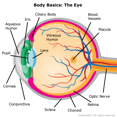
Vision is the process by which images captured by the eye are interpreted by the brain, and the visible part of the eye is where the process of sight begins. On the front surface of the eye is the see-through, circle-shaped cornea. You can't see a person's cornea the way you can see the colored part of the eye behind it — the cornea is like a clear window that focuses light into the eye.
Behind the cornea is a watery fluid called the aqueous humor. The cornea and aqueous humor form an outer lens that refracts (bends) light on its way into the eye. This is where most of the eye's focusing work is done.
The colored circular membrane in the eye just behind the cornea is called the iris. The iris controls the amount of light entering the eye through the pupil, which is the opening in the center of the iris that looks like a tiny black circle.
Like a camera, which controls the amount of light coming in to prevent both overexposure and underexposure, the iris becomes wider and narrower, changing the size of the pupil to control the amount of light entering the eye. The pupil gets bigger when more light is needed to see better and smaller when there's plenty of light.
The eye's lens sits just behind the iris. Just like a camera lens, the eye's lens focuses light to form sharp, clear images. Light that has been focused through the cornea and aqueous humor hits the lens, which then focuses it further, sending the light rays through the vitreous humor and onto the retina.
To focus on objects clearly at varying distances, the eye's lens needs to change shape. The ciliary (pronounced: SIL-ee-air-ee) body contains the muscular structure in the eye that changes the shape of the eye's lens. In people who have normal vision, the ciliary body flattens the lens enough to bring objects into focus at a distance of 20 feet or more. To see closer objects, this muscle contracts to thicken the lens. Young children can see objects at very close range; many people over 45 have to hold objects farther and farther away to see them clearly. This is because the lens becomes less elastic with age.
The retina (the soft, light-sensitive layer of tissue that lines the back of the eyeball wall) is made up of millions of light receptors called rods and cones. Rods are much more sensitive to light than cones. Each eye has about 120 million rods that help us see in dim light and detect shades of gray, but they cannot distinguish colors. In comparison, the 6 million cones in each eye allow us to see in bright light and they also sense color and detail.
The macula (pronounced: MAK-yuh-luh) is a small, specialized area on the retina that helps the eyes see fine details when we look directly at an object. It contains mainly cones and few rods.
When focused light is projected onto the retina, it stimulates the rods and cones. The retina then sends nerve signals are sent through the back of the eye to the optic nerve. The optic nerve carries these signals to the brain, which interprets them as visual images. The portion of the brain that processes visual input and interprets the messages that the eye sends is called the visual cortex.
As in a camera, the eye's lens transmits light patterns upside down. The brain learns that the impulses received from the upper part of the retina are really from the lower part of the object we're seeing and vice versa.
Most people use both eyes to see an object. This is called binocular vision, and images are formed on the retina of each eye. These images are slightly different because the object is being viewed from slightly different angles. Nerve signals representing each image are sent to the brain, where they are interpreted as two views of the same object. Some of the nerve fibers from each eye cross, so each side of the brain receives messages from both eyes. Through experience, the brain learns to judge the distance of an object by the degree of difference in the images it receives from the two eyes. This ability to sense distance is called depth perception.
What Causes Vision Problems?
Vision is a fine-tuned process. All the parts of the eye — and the brain — need to work together so a person can see correctly. Because the eye's structure is so complex, though, a lot of things can go wrong.
Some of the most common eye problems are refractive errors. These are the problems that eye doctors check for routinely in a vision test. Refraction means bending of light rays to focus the light coming from an image. Refractive errors are problems with the focusing of the eye, because of the way the eye is shaped, which causes the image you see to be blurred.
Refractive errors include:
Astigmatism. In astigmatism (pronounced: uh-STIG-muh-tih-zum), there's a problem with the curve of the cornea. This causes part of the eye's image to be blurry. Corrective lenses such as contact lenses or glasses can usually correct vision in people with astigmatism.
Myopia. Also called nearsightedness or shortsightedness, myopia (pronounced: my-OP-ee-uh) happens when the eye focuses the image of an object in front of the retina instead of directly on it. In most cases, people can't see well far away, but can see objects clearly close up. The condition tends to get somewhat worse through childhood and adolescence, but stabilizes in adulthood. People with this condition may need to wear glasses or contacts to correct their vision. Laser eye surgery is sometimes used in adults to correct nearsightedness permanently by changing the shape of the cornea. Laser surgery is not used for teens because the eye may still be growing and the refractive error changing.
Hyperopia. Also called farsightedness or longsightedness, hyperopia (pronounced: hy-per-OP-ee-uh) happens when the incoming image is not focused on the retina, but behind it. This may make it difficult to see close objects clearly, with far-off objects seen more easily. Many younger children are hyperopic, but because of the ability of the eye to focus itself, may not need glasses to correct this. Glasses or contact lenses can correct this problem in kids and teens when needed. Most adults develop a form of farsightedness called presbyopia as they get older.