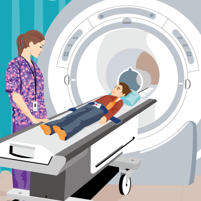
Magnetic Resonance Imaging (MRI): Brain
What's an MRI (Magnetic Resonance Imaging)?
An MRI (magnetic resonance imaging) is a safe and painless test that uses magnets and radio waves to make detailed pictures of the body's organs, muscles, soft tissues, and structures. Unlike a CAT scan, an MRI doesn’t use radiation.
MRIs are done in hospitals and at radiology centers.
What Is a Brain MRI?
A brain MRI produces detailed pictures of the brain and the brain stem.
Why Are Brain MRIs Done?
A brain MRI can help doctors look for conditions such as bleeding, swelling, problems with the way the brain developed, tumors, infections, inflammation, damage from an injury or a stroke, or problems with the blood vessels.
The MRI also can help doctors look for causes of headaches or seizures. It can show if a shunt is working.
In some cases, MRI can provide clear images of parts of the brain that can't be seen as well with an X-ray, CAT scan, or ultrasound, making it particularly valuable for finding problems with the pituitary gland and brain stem.
What if I Have Questions?
If you have questions about the brain MRI or the results of the test, speak with your doctor. You can also talk to the MRI technician before the exam.


© 1995- The Nemours Foundation. KidsHealth® is a registered trademark of The Nemours Foundation. All rights reserved.
Images sourced by The Nemours Foundation and Getty Images.
