X-Ray Exam: Scoliosis
What's an X-Ray?
An X-ray is a safe and painless test that uses a small amount of radiation to make an image of bones, organs, and other parts of the body.
The X-ray image is black and white. Dense body parts, such as bones, block the passage of the X-ray beam through the body. These look white on the X-ray image. Softer body tissues, such as the skin and muscles, allow the X-ray beams to pass through them. They look darker on the image.
X-rays are commonly done in doctors’ offices, radiology departments, imaging centers, and dentists’ offices.
What Is a Scoliosis X-Ray?
A scoliosis X-ray creates detailed images of the spine. An X-ray machine sends a beam of radiation through the back, and an image is recorded on a computer or special film. The scoliosis X-ray includes the thoracic spine (upper back) and the lumbar spine (lower back).
X-rays are done by an X-ray technician in the radiology department of a hospital, a freestanding radiology center, or a health care provider's office. They usually take two pictures of the spine:
- one from the back (posteroanterior or PA view)
- one from the side (lateral view)
Sometimes other views are taken as the person bends to the side.
Why Are Scoliosis X-Rays Done?
Children with scoliosis have a spine that curves from side to side, like an S or a C. Small curves generally don't cause problems, but bigger curves can be visible and cause discomfort. A curved spine can cause the trunk to shift to the left or right. One shoulder blade may be higher than the other, or the waist may be uneven, with a tendency to lean to one side.
A severe curve can even affect breathing and heart function and lead to damage in the joints of the spine and pain during adulthood.
Primary care providers routinely check kids for scoliosis during regular checkups. If scoliosis is suspected, they may order X-rays to measure the curvature of the spine. The angle of the curve, measured in degrees on the X-ray, will help them decide whether it needs to be treated and, if so, how.
X-rays also help determine the type of scoliosis and how mature the child's skeleton is, which helps the provider predict if the curve might get worse. The scoliosis X-ray might be repeated at regular intervals (often every 6 months) to check whether the curve is getting bigger or to monitor the effects of treatment.
The provider might refer your child to a specialist for further evaluation. If a neurological problem is suspected, the doctor may order a magnetic resonance imaging (MRI) study to look at the spinal cord.
What Happens During a Scoliosis X-Ray?
Although the procedure may take about 10 minutes, actual exposure to radiation is usually less than a few seconds.
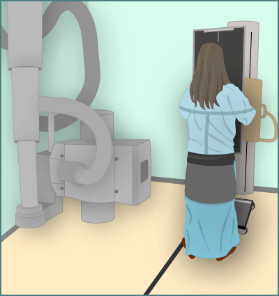
Traditional X-Ray
Your child will go into a special room with a table and a large X-ray machine hanging from the ceiling or wall. Parents usually can stay with their child to provide reassurance. If you stay in the room while the X-ray is done, you'll wear a lead apron to protect certain parts of the body. The child's reproductive organs also are protected with a lead shield.
A scoliosis X-ray is usually done with the child in a standing position. The technician will position the child, then step behind a wall or into an adjoining room to operate the machine. Two X-rays are usually taken (from the back and from the side), so the technician will return to reposition the child for each X-ray. Sometimes doctors will ask for more views.
Older kids will be asked to hold their breath and remain still for 2–3 seconds while each X-ray is taken. Infants may require gentle restraint. Keeping still is important to prevent blurring of the X-ray image.
EOS Imaging System
Some scoliosis X-rays are done with an EOS X-ray machine. This machine uses a very low dose of radiation, which is ideal in scoliosis patients who need to get X-rays regularly.
The child goes into a small booth, where both the picture from the front and the sides are taken at the same time. The X-rays are done in less than 30 seconds. Your child won't need to wear a lead apron due to the much lower doses of radiation.
What If I Have Questions?
If you have questions about the scoliosis X-ray, speak with your doctor. You can also talk to the X-ray technician before the procedure.
 Note: All information is for educational purposes only. For specific medical advice,
diagnoses, and treatment, consult your doctor.
Note: All information is for educational purposes only. For specific medical advice,
diagnoses, and treatment, consult your doctor.
© 1995- The Nemours Foundation. KidsHealth® is a registered trademark of The Nemours Foundation. All rights reserved.
Images sourced by The Nemours Foundation and Getty Images.