


X-Ray Exam: Upper Leg (Femur)
What's an X-Ray?
An X-ray is a safe and painless test that uses a small amount of radiation to make an image of bones, organs, and other parts of the body.
The X-ray image is black and white. Dense body parts, such as bones, block the passage of the X-ray beam through the body. These look white on the X-ray image. Softer body tissues, such as the skin and muscles, allow the X-ray beams to pass through them. They look darker on the image.
X-rays are commonly done in doctors’ offices, radiology departments, imaging centers, and dentists’ offices.
What's a Femur X-Ray?
In a femur X-ray, an X-ray machine sends a beam of radiation through the upper leg (the area between the hip and knee), and an image is recorded on special film or a computer. This image shows the soft tissues and the bone in the upper leg, which is called the femur (FEE-mer).
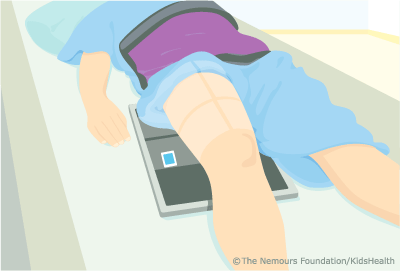
An X-ray technician will take pictures of the femur:
- from the front (AP, or anteroposterior view)
- from the side (lateral view)
Femur X-rays are done with the person lying down or standing. They should stay still for 2-3 seconds while each X-ray is taken so the images are clear. If an image is blurred, the X-ray technician might take another one.
Why Are Femur X-Rays Done?
A femur X-ray can help doctors find the cause of pain, limping, tenderness, swelling, or deformity of the upper leg. It can show a broken bone, and after a broken bone has been set, it can show if the bone is aligned and if it has healed properly.
An X-ray can help doctors plan surgery, when needed, and check the results after it. It also can help detect cysts, tumors, or other diseases in the bone, including later stages of bone infections.
What if I Have Questions?
If you have questions about the femur X-ray or what the results mean, talk to your doctor.
 Note: All information is for educational purposes only. For specific medical advice,
diagnoses, and treatment, consult your doctor.
Note: All information is for educational purposes only. For specific medical advice,
diagnoses, and treatment, consult your doctor.
© 1995- The Nemours Foundation. KidsHealth® is a registered trademark of The Nemours Foundation. All rights reserved.
Images sourced by The Nemours Foundation and Getty Images.